The spectre of Bond, fusiform gyrus and ‘are faces special?’

Last night, I went to see “Spectre”, the new Bond movie, in the O2 centre on Finchley Road.

A review of it, in the New Yorker, is here.

Being careful not to do any ‘spoilers’, towards the end of a rather long but excellent film, there is a futuristic scene involving James Bond. The Bond baddie of this movie mentions ‘the fusiform gyrus’, which is indeed an interesting part of the brain.

Fans of cognitive neurology will of course recognise this part of the brain at once.

The temporal lobe, part of the brain near the ear, has a number of different functions including learning and memory. That’s why when, early on in the disease process of Alzheimer’s disease affecting the ‘hippocampus’ in the temporal lobe, persons often remark losing memory of whole recent events. Elsewhere in the temporal lobe is a distinct perceptual function – in particular for the higher order complex processing of objects. It’s been known for over thirty years now that there are neurones in the ‘superior temporal sulcus’ which appear particularly sensitive to faces.

One of the classic reports in the literature is here.

The superior temporal sulcus and fusiform gyrus are both parts of the human brain which are intimately connected in both structure and function. The question for future research will be to find out why and how.

The idea that a part of the brain has a particular function has been around for some time ago.

Phrenology (from Greek φρήν (phrēn), meaning “mind”, and λόγος (logos), meaning “knowledge”) is a pseudoscience which was primarily focused on measurements of the human skull, based on the concept that the brain is the organ of the mind, and that certain brain areas have localised, specific functions or modules (source).
 PhrenologyPix
All this came to a fore in a classic from the MIT Press from Jerry Fodor, “The Modularity of Mind” (1983).
It is of course a hugely powerful issue whether one part of the brain holds the key to a particular function. But it’s been latterly been realised that localised parts of the brain ‘doing function’ identified in a simplistic way, say the Wernicke area for language comprehension, may in fact be a rather complicated discrete distributed neural network.
An example of a recent paper from the cognitive neurology literature is here.
But are faces special?
This was indeed a hot enough question for the great Oliver Sacks to devote a piece of work to, entitled “The man who mistook his wife for a hat”.


That face perception in humans was truly recognised in Bodamer’s 1947 classic paper on prosopagnosia.

Problems in perception of face identity and facial expression can occur in isolation. Though there are methodological concerns, it could be that certain persons living with Huntington’s disease could have a real problem, for example, with the perception of disgust.

Dementia, the umbrella term usually given for chronic progressive diseases of brain, can affect any function – it’s not just about memory. Therefore unsurprisingly persons with dementia might theoretically have problems with face perception.

One intriguing example is the “Capgras delusion“. The Capgras delusion (or Capgras syndrome) describes where a person holds a delusion that a friend, spouse, parent, or other close family member (or pet) has been replaced by an identical-looking impostor.

There are various approaches which can be tried here perhaps.

One which is interesting is “entering the reality”, as described here.

capgras

This is part of the ethical debate about ‘therapeutic lying’ which was introduced last week in an excellent plenary session by Toby Williamson at the UK Dementia Congress 2015 in Telford.

Of course, entering the reality of James Bond is an altogether different matter.

Decisions in different types of dementia

Decisions are fundamental to our lives.

Decision making is a fundamental and complex skill which is crucial at any age. We all have to face decisions regarding their health care, medical treatment, retirement, housing, transport, and finances, for example.

We not only have to consider the benefit of a decision for their current living situation, but also to anticipate the consequences of decisions of such actions in the nearer and farer future. We need to hold the decision in my memory for long enough to think through strategically the options, and be able to action an outcome.

Everyday life requires numerous and fast decisions. Often these decisions have an uncertain result. Wrong decisions may thus have severe consequences in several domains.

Disturbances in an ability to make decisions or to anticipate the possible consequences of decisions can result in massive problems.

In the last decade in cognitive neuroscience and cognitive neurology, there has been an increasing interest to investigate neural basis of decision-making abilities and disturbances both in healthy subjects and to people where there has been some disruption.

But we have been able to build up a coherent picture of this using neuropsychological and neuroimaging techniques.

An assessment of cognitive deficits in neurodegenerative diseases has focused so far almost entirely on memory, language, attention, visuospatial perception and executive functioning (Gleichgerrcht et al., 2010).

In the past decade, however, the study of decision-making in these conditions has increased, prompting the development of new tasks that have enabled this cognitive process to be readily assessed. In clinical practice, it is not uncommon to find early persons with behavioral-variant frontotemporal dementia (bvFTD) who, to a considerable extent, are intellectually unimpaired, while relatives and caregivers depict a strikingly different picture: they claim that these patients show severe changes in their behaviour and real-life decision-making skills (e.g. Rahman et al., 1999; Manes et al., 2011).

The literature has so able to identify the orbitofrontal, anterior cingulate, and dorsolateral prefrontal cortices as being critical to decision-making (Rosenbloom, Schmahmann and Price, 2012).

A schematic view of the important neural substrates proposed by Rahman and colleagues (Rahman et al., 2001) is shown in Figure 1.

TICS

Rahman and colleagues showed that patients with behavioural variant frontemporal dementia exhibited a profile of risk-taking, not impulsive, behaviour in decision-making, suggestive of dysfunction in the ventromedial prefrontal or orbitofrontal cortex. Kloeters and colleagues (Kloeters et al., 2013), fourteen years later, published results showing that atrophy in the orbitofrontal cortex and amygdala correlated with performance on the Iowa Gambling Task used in their study to examine decision-making.

A large proportion of human cognitive social neuroscience research has focused on the issue of decision-making thus far.

Impaired decision-making is a symptomatic feature of a number of neurodegenerative diseases, but the nature of these decision-making deficits depends on the particular disease.

Once you’ve met one person with dementia, you’ve met one person with dementia. Each person with dementia will have a cognitive profile according how far progressed the condition has reached, and the extent to which functional problems are perceived. This might depend on the likely diagnostic category in which a patient living with dementia finds himself or herself.

Examining the qualitative differences in decision-making impairments associated with different neurodegenerative diseases provides potentially valuable information regarding the underlying neural basis of decision-making.

A good account of decision-making in different neurological conditions including dementia is provided by Brand, Labudda and Markowitsch (2006).

Figure 2 shows a schematic view of some of the key processes.


Final picture for ch 13

The features of their model are as follows.

General problem solving strategies, also stored in long-term memory, need to be recalled in order to evaluate which strategy seems to be appropriate in order to decide advantageously. The recall of this information, including personal autobiographical experiences and general strategies that have been developed during life, is triggered and controlled by executive components, for example cognitive flexibility.

In working memory, the features of the current decision and the retrieved information from long-term memory are combined to generate or initiate a current decision strategy that guides the decision.

In this process, “somatic markers”, which means biasing signals from the body or mental representations of them, can also guide the selection of an appropriate strategy. The decision itself leads to positive or negative feedback (e.g., gain or loss of a specific amount) that activates an bodily autonomic response.

The feedback – or the somatic markers, which are the results of the emotional feedback – can also result in an alteration of the information stored in long-term memory as well as – in a more direct way – the representation of somatic markers associated with comparable decisions.

The comparison of the profiles of decision making in different conditions, which can cause dementia, are arguably helpful in predicting what the person with dementia might expect. Several studies have reported altered decision-making in Parkinsons’s disease (Perretta et al., 2005) and pathological gambling has been found in Parkinsons’s disease patients with L-Dopa medication (Weintraub et al., 2006) attributing a key role to the chemical dopamine in taking risky decisions.

Recent studies also investigated decision-making in Huntington’s disease and found that learning and memory processes, rather than motivational processes, are responsible for decision-making deficits in this group (Busemeyer and Stout, 2002).

Hampton and O’Flaherty (2007, some years ago, mapped out the neural substrates of reward-related decision making with functional MRI. They identified that the combined signals from three specific brain areas (anterior cingulate cortex, medial prefrontal cortex, and ventral striatum) were found to provide all of the information sufficient to decode subjects’ decisions out of all of the regions studied.

These findings appear to implicate a specific network of regions in encoding information relevant to subsequent behavioral choice. Evidence for the important role of the orbitofrontal cortex and the amygdala in decision-making particularly under ambiguous conditions comes from a recent study by Hsu and colleagues (Hsu et al., 2005)).

Dementia of the Alzheimer type (DAT), the cause of the most cases of dementia worldwide, is typically characterised by typical structural, neurochemical and cognitive changes as the disease progresses.

Pathological changes in mild DAT affect primarily the medial temporal lobes and limbic structures (e.g., entorhinal cortex, hippocampus), and then extend to the association cortices of the frontal, temporal and parietal lobes (Braak and Braak, 1991).

Ha and colleagues have argued that the changes in DAT fundamentally alter the frames of reference for making decisions (Ha et al., 2012).

The study by Delazer and colleagues further highlighted important differences in decision-making between mild DAT patients and healthy controls (Delazer et al., 2007). Findings from the study by Sinz and colleagues are consistent with the notion that decisions under ambiguity as well as decisions under risk are impaired in mild DAT (Sinz et al., 2008). It may thus be expected that patients with mild DAT have difficulties in taking decisions in everyday life situations, both in cases of ambiguity (information on probability is missing or conflicting, and the expected utility of the different options is incalculable) and in cases of risk (outcomes can be predicted by well-defined or estimable probabilities).

The legal instrument to assess capacity through the Mental Capacity Act (2005) is very blunt. Characterising an ability of a person living with dementia to make optimal decisions is essential for giving confidence to that person (and those closest to him and her) that such risks are being managed appropriately.

It is likely that the implementation of the Mental Capacity Act will come under increasing scrutiny, in parallel with advances in decision-making research in cognitive neuroscience and cognitive neurology.


References

Braak, H., Braak, E. (1991) Neuropathological staging of Alzheimer-related changes, Acta Neuropathologica (Berl), 82, pp. 239–259.

Brand, M., Labudda, K., Markowitsch, H.J. (2006) Neuropsychological correlates of decision-making in ambiguous and risky situations, Neural Netw, 19(8), pp. 1266-76.

Busemeyer, J. R., Stout, J. C. (2002) A contribution of cognitive decision models to clinical assessment: Decomposing performance on the Bechara gambling task, Psychological Assessment, 14, pp. 253–262.

Delazer, M., Sinz, H., Zamarian, L., Benke, T. (2007) Decision-making with explicit and stable rules in mild Alzheimer’s disease, Neuropsychologia, 45(8), pp. 1632-41.

Gleichgerrcht, E., Ibáñez, A., Roca, M., Torralva, T., Manes, F. (2010) Decision-making cognition in neurodegenerative diseases, Nat Rev Neurol, 6(11), pp. 611-23.

Ha, J., Kim, E.J., Lim, S., Shin, D.W., Kang, Y.J., Bae, S.M., Yoon, H.K., Oh, K.S. (2012) Altered risk-aversion and risk-taking behaviour in patients with Alzheimer’s disease, Psychogeriatrics, 12(3), pp. 151-8.

Hampton, A.N., O’Doherty, J.P. (2007) Decoding the neural substrates of reward-related decision making with functional MRI, Proc Natl Acad Sci U S A, 104(4), pp. 1377-82.

Hsu, M., Bhatt, M., Adolphs, R., Tranel, D., Camerer, C. F. (2005) Neural systems responding to degrees of uncertainty in human decision-making, Science, 310, pp. 1680–1683.

Kloeters, S., Bertoux, M., O’Callaghan, C., Hodges, J.R., Hornberger, M. (2013) Money for nothing – Atrophy correlates of gambling decision making in behavioural variant frontotemporal dementia and Alzheimer’s disease, Neuroimage Clin, 2, pp. 263-72.

Manes, F., Torralva, T., Ibáñez, A., Roca, M., Bekinschtein, T., Gleichgerrcht, E. (2011) Decision-making in frontotemporal dementia: clinical, theoretical and legal implications, Dement Geriatr Cogn Disord, 32(1), pp. 11-7.

Rahman, S., Sahakian, B.J., Hodges, J.R., Rogers, R.D., Robbins, T.W. (1999) Specific cognitive deficits in mild frontal variant frontotemporal dementia, Brain, 1999, 122 (Pt 8), pp. 1469-93.

Rosenbloom, M.H., Schmahmann, J.D., Price, B.H. (2012) The functional neuroanatomy of decision making, J Neuropsychiatry Clin Neurosci, 24(3), pp. 266-77.

Sinz, H., Zamarian, L., Benke, T., Wenning, G.K., Delazer, M. (2008) Impact of ambiguity and risk on decision making in mild Alzheimer’s disease, Neuropsychologia, 46(7), pp. 2043-55.

Weintraub, D., Siderowf, A. D., Potenza, M. N., Goveas, J., Morales, K. H., Duda, J. E., Moberg PJ, Stern MB. (2006) Association of dopamine agonist use with impulse control disorders in Parkinson disease, Archives of Neurology, 63, pp. 969–973.


Awareness about dementia is not just public ignorance: it’s also critical to living with dementia

Often I’m struck about how the ‘awareness’ focus in dementia is making people in general public simply knowledgeable that dementia exists in 800,000 people in the UK.

But awareness about symptoms in persons living with dementia themselves is also a critical component, and cannot be factored out of the debate in current policy drive to identify the missing people undiagnosed dementia.

Policy wonks without a scientific or clinical training in dementia have become very adept at blaming GPs for underdiagnosis of dementia, but people who have some knowledge of this specialist field know that the situation is far more complicated. Other issues include perhaps a reluctance of people to seek a diagnosis because of the life-changing impact that such a diagnosis might make. There may also be nuances between different ethnic or social groups in society which might act as ‘barriers to diagnosis’. Also, some persons with dementia may be genuinely unaware of the extent of their own symptoms.

To be fair, it’s impossible for anyone who doesn’t have a diagnosis of a dementia to understand completely what living with dementia really means. Norman McNamara, who was diagnosed with dementia a few years ago at the age of fifty comments: “What can be worse than having dementia?” “It’s knowing you have dementia – it’s like having two diseases, having it, and knowing you have it.”

This is a helpful description of ‘insight’, that people with dementia can have into their own conditions. In this video, Norman reports symptoms which he knows are getting worse, and which knows are visibly getting worse to his wife, Elaine. Patients with neurological disorders are often partially or completely unaware of the deficits caused by their disease. This impairment is referred to as “anosognosia”, and it is very common in neurodegenerative disease, particularly in frontotemporal dementia.

The mechanisms underlying this phenomenon are generally poorly understood. It’s likely, however, memory for facts and events likely plays an important role. In addition, the frontal lobe systems are important for intact self-awareness, but the most relevant frontal functions have not been identified. Motivation required to engage in self-monitoring and emotional activation marking errors as significant are often-overlooked aspects of performance monitoring that may underlie anosognosia in some patients.

Another common type of dementia is a behavioral variant frontotemporal dementia (bvFTD), characterised by a slow change in personality and behaviour, is often unnoticed by the individual himself or herself. Loss of insight is a prominent clinical manifestation of this condition, but its characteristics are poorly understood. Indeed, Mario Mendez and Jill Shapira reported in 2005 some research into what appeared to cause this lack of insight in this particular condition. They found that it is associated with low blood flows in the right hemisphere, particularly the frontal lobe, the part of the brain near the front of the head.

For the most common type of dementia, the dementia of the Alzheimer type, the generally widely-held belief is that persons experience a progressive loss of insight as the severity of dementia increases. People with this type of dementia can get particularly forgetful. Most people aren’t fully aware of their impaired abilities, which doctors describe as a “lack of insight”. This can put them at risk of injury from unsafe actions and also make them less willing to seek and comply with treatment.

However, understanding a person’s level of insight can help doctors and carers better manage their treatment and daily needs, but gauging insight can be difficult. The usual approach is to ask patients questions about their current abilities and compare their answers with those from an ‘informant’, which is usually a family member or someone else close to the patient.

But this method isn’t ideal, as it relies on the informant’s opinion of the patient’s abilities, which can be swayed by factors such as how well they know the patient and how distressing they find their behaviour.

Norman often states that ‘once you’ve met one person with dementia, you’ve met one person with dementia’. This means that for any one person with dementia there’ll be different extents of symptoms of illness, different extents of abilities, different levels of insight, and therefore different perceptions of ‘living well with dementia’. So, it is arguably difficult to compare whether one type of dementia is ‘worse’ than other?

Dr Mitul Mehta from the Institute of Psychiatry asks for greater scrutiny for what the Cambridge Cognition test for Alzheimer’s Disease actually examines

Dr Mitul Mehta heads up the neuropharmacology group at the Institute of Psychiatry.

Here, he describes the Cambridge Cognition “paired associates learning test”, where you have to remember which boxes contain particular objects on a screen.

Both Dr Mehta and I both worked in the research laboratory in Cambridge which did research into this test at the end of the 1990s.

It might be sensitive to the memory deficits in people with early Alzheimer’s disease, and indeed was cited by David Cameron in his G8 dementia summit speech.

But what specifically is impaired is difficult to work out, because the test measures more than one thing.

There is an attentional component. We know through a number of routes, in whatever way you decide to investigate variants of this task, the brain regions activated tend to encompass those at the front and back of the back/side (the “frontoparietal network”).

It also loads heavily on working memory.

But these are not areas thought by most to be involved early in Alzheimer’s disease.

But it also loads heavily on episodic memory and learning. If we could show better that it’s this learning component that’s affected, this would make interpretation of the test far easier.

That this test also picks up memory problems in ‘mild cognitive impairment’, which is not Alzheimer’s disease, is a problem. The question
is how many of these people who have mild cognitive impairment, who don’t have Alzheimer’s disease, get told that they may have Alzheimer’s disease, and go onto receive further investigations which might include a specialist brain scan.

There is, there, a legitimate question to be asked about which parts of the brain are activated there.

There has never been robust evidence, because of the way in which this task has been investigated using a brain scanner, that it is specifically the learning components that activate certain parts of the brain, known as the parahhippocampal gyrus and entorhinal cortex.

It is these parts of the brain which ‘gate information’ into the hippoocampus proper, in the temporal lobe. That’s the part of the brain by your ear.

bilateral hippocampus

If the task could be properly neuroimaged to tease out this learning component, we’d be much further forward. For what it’s worth, I think the task will activate the parahippocampus gyrus and perirhinal cortex, but it really is a question of showing this properly. This I feel has yet to be done.

Concerns about the paired associates learning test for dementia

To begin to understand how a cathode-ray TV set works, I could remove one component called the “transistor”, and the picture disappears. It would be an incorrect conclusion to say that the purpose of that transistor is to produce the picture. However, I could argue correctly that the transistor was somehow part of the system required to produce the picture.

If I showed the transistor was particularly “hot” while the TV set was on, producing a picture, it might be reasonable for me to conclude the transistor was involved in producing the picture.

This is the sort of basic approach still used to work out what is going on in the brains and minds of people with Alzheimer’s disease, typical presentations of which might be memory problems. You can see whether removing parts of the brain in humans produces similar effects to the problems in thinking found in Alzheimer’s disease. Or alternatively, you could just try to look at the system of components in the brain which might be contributing to memory in brains working normally.

TV set

Whatever, it’s a puzzle. In this particular case, it’s a puzzle to solve correctly.

An innovation culture in the diagnosis of Alzheimer’s disease

David Cameron praised Cambridge Cognition’s work in developing new innovative tests for Alzheimer’s disease in the G8 summit held towards the end of last year.

There has been concern that some individuals with Alzheimer’s disease do not receive their diagnoses in a particularly fast way. A number of explanations for this have been offered, including medical personnel not being able to spot the symptoms of Alzheimer’s disease easily.

It is also helpful to understand what an “innovation” is. An innovation might be a product which enables you do something much more easily, and depends for its success popular uptake by the user. Strictly speaking, paper was an innovation too. However, the rise in cost of diagnosing Alzheimer’s disease, arguably, is an intriguing example of “Baumol’s cost disease“.

Individuals with Alzheimer’s disease have memory problems which are typically not thought to be qualitatively similar to those found in ageing elderly individuals. Often such people have real problems in navigating around environments. It is clearly a very laudable aim to have a bedside test which might be able to alert a physician to an underlying memory problem in Alzheimer’s disease.

The benefits and concerns, and my passing involvement

There are a number of important caveats here. Not all dementias are Alzheimer’s disease. There are in fact hundreds of dementias, some of which are reversible. Whatever test is used, the test should be sensitive enough to identify reliably a genuine thinking problem in Alzheimer’s disease, but should not be so ‘broad brush’ the test also misattributes memory problems, say found in the ‘mild cognitive impairment’ or even depression, to Alzheimer’s disease. Such mislabelling can perceivably cause distress, and cause people to be caught up in the medical system for further lengthy tests when they should not have been in the first place. On the other hand, it is of concern that the diagnosis might be missed in some people, and hence the drive from the Department of Health and the Alzheimer’s Society in “The Prime Minister’s Dementia Challenge”.

I wish Cambridge Cognition well, not least because I have worked with CANTAB whilst a graduate student at the University of Cambridge. In fact, some of my papers are cited in their bibliography. Their search facility is here.

Bibliography

The CANTABmobile “paired associates learning” test

To explain the “paired associates learning” test from first principles, and I’m not using actual screenshots, imagine me presenting you with a number of blank boxes dotted around the screen.

Fig 1

And I open each box in turn and reveal a shape to you. I can present the problem with a varying number of shapes.

Fig 3     Fig 2

After showing you all the shapes, I then present to you a shape and ask you to identify the box in which it was first presented.

Fig 4

Cambridge Cognition in welcoming the Draft National Plan to Address Alzheimer’s disease in my opinion set out entirely correctly the advantages of this computerised testing battery; including fast, not culturally biased, not heavily loading on language, norm-referenced, culturally unbiased, and easy-to-use.

The reasoning behind it being sensitive to early Alzheimer’s disease – but what about mild cognitive impairment?

To understand why the narrative for the test being so attractive in early Alzheimer’s disease, you have to understand that this test has been found to be sensitive to functions of particular brain areas. If you chop out bits of the brain near the front of the head (frontal cortex) or near the ear (temporal cortex), performance on this task is impaired, as Prof Adrian Owen showed when he was a post-doctoral fellow (paper here). With hindsight, perhaps Owen should have looked at the effects of other brain areas further back in the brain, such as the parietal cortex, which are also now thought to be important in memory for spatial cues.

A consistent finding has been loss of brain cells in the “entorhinal cortex”, in the temporal cortex, early in Alzheimer’s disease (see for example here). Therefore, that the paired associates learning test should identify memory problems in early Alzheimer’s disease immediately makes intuitive sense.

But the issues I feel are much more complicated, and I wish Cambridge Cognition well in clarifying them.

If it’s not Alzheimer’s disease, what else could be causing the memory problems?

One possibility is “mild cognitive impairment”. It is described, for example on the authoritative Mayo Clinic website, that:

“Mild cognitive impairment (MCI) is an intermediate stage between the expected cognitive decline of normal aging and the more serious decline of dementia. It can involve problems with memory, language, thinking and judgment that are greater than normal age-related changes. If you have mild cognitive impairment, you may be aware that your memory or mental function has “slipped.””

David Hart, Senior Business Development Manager of Cambridge Cognition, kindly sent Dr Peter Gordon the rationale for the use of the CANTAB task by Dr Andrew Blackwell, their Chief Scientific Officer (as produced on Peter’s blog here).

Cambridge Cognition concede that distinguishing between MCI and Alzheimer’s Disease “is difficult”, but this is a distinction that must be arrived at otherwise a test potentially will give “false positives” – but no test is perfection, and it basically is impossible to strive for perfection. What we all trying avoid is where a test for possible dementia itself is expensive followed by a further expensive investigation to show the original result was a false positive – or as the Express euphemistically called it recently, “Dementia diagnosis proved wrong by new super scanner”.  (It is important to state clearly here that no details are given how a diagnosis had been arrived at previously for Ros Davies.)

To give them credit, Cambridge Cognition cite the Chandler et al. (2008) paper, but the full citation of this is “Alzheimer’s & Dementia: The Journal of the Alzheimer’s Association Volume 4, Issue 4, Supplement , Pages T551-T552, July 2008″ – i.e. it is a supplement of abstracts not full papers. This particular abstract can be viewed here.

It is hoped that this full study will have been published elsewhere, and if so Cambridge Cognition will need to update their website with the full paper. Notwithstanding this, the numbers of individuals in each group are disappointingly low: there are seventeen with putative MCI and twelve with putative Alzheimer’s disease.

Is this task actually sensitive and specific?

However, the discussion by Dr Andrew Blackwell and colleagues in his 2004 paper is useful. I have more than a passing interest in that paper as the main author on that paper was one of my PhD supervisors at Cambridge, Prof John Hodges. John has also kindly written one of my three Forewords for my book, “Living well with dementia” to be published on January 14th 2014.

Blackwell remarks correctly that this task has been used to distinguish between unipolar depression and Alzheimer’s disease in Rachel Swainson’s study. But is this enough? I looked to the previous Beats study in “geriatric depressive”, and there was nothing forthcoming there. How confident can one be that only early patients with Alzheimer’s disease, and not those severely depressed or with an underactive thyroid, will perform abnormally on the PAL? Personally, I’m not at all confident yet, despite the Swainson study, but these fears can easily be allayed with a sensitivity/specificity study of much higher power.

Blackwell is however correct in citing my study with Dr Andy Lee in that patients with semantic dementia and behavioural variant frontotemporal dementia are relatively unimpaired, though the clinical presentations of the frontotemporal dementias can be quite clearly different in the clinic from Alzheimer’s disease. Completing the double dissociation, I did find that the behavioural variant of frontotemporal dementia did present with rather specific risk-taking decision-making of its own.

But in the meantime the comparison with frontotemporal dementias is useful.

Lee

Nonetheless, this approach is being rolled out.

On 28 June 2013, the use of CANTABmobile was described as follows:

“The Guildford and Waverley Clinical Commissioning Group (CCG) is leading the use of an innovative new iPad-based memory assessment  system as part of a national push to decrease dementia diagnosis waiting times and streamline the referral process.   Accessed through NHS medical professionals, CANTABmobile enables GPs to test a patient’s episodic memory through an easy to use and administer 10-minute cognitive assessment.”

The CANTAB paired associates learning test is pictured under the heading “intuitive touchscreen interface”. if you go to “download information” on this page.

It was covered in the national media here: for example Victoria MacDonald’s report (this page provides a criticism of another report by Victoria MacDonald this time over Prof Brian Jarman’s proposed HSMR data by NHS Consultant, Dr Jacky Davis).

So what does this task test?

In understanding how the task works in reality, I found a paper where Prof Ed Bullmore and colleagues put individuals with Alzheimer’s disease and control subjects performing the task into a scanner really helpful.  Bullmore and colleagues frontloaded their discussion with the following comment:

“Independent of the level of difficulty, the majority of subjects in both groups activated a network of brain regions, including the anterior cingulate, lateral, and medial occipitoparietal and frontal cortices, during successful encoding and retrieval.”

This is interesting as it doesn’t point to the usual suspects of the narrative, i.e. the entorhinal cortex and other parts of temporal lobe. Even Andrew Blackwell had described how the damage to the entorhinal cortex might possibly account dor deficits on the paired associates task:

“The transentorhinal region is a complex transitional area located between the entorhinal region proper and the adjacent temporal isocortex. It has been suggested that damage to this site in early [Alzheimer’s disease] disrupts reciprocal connections with the hippocampal formation and that this disruption underlies deficits in episodic memory.”

But on reflection is this wholly a surprise? Ed Bullmore and colleagues from their results, also from Cambridge, discuss that the lateral parietal activations reported during episodic memory tasks are thought to reflect recognition processes and retrieval processing of spatial information. Medial parietal activity has been proposed to underlie imagery and retrieval success.

I don’t feel it’s altogether surprising given what is known about the build-up of pathology in Alzheimer’s disease, either. The authors of one study looking at this report that:

“[18F]FDDNP-PET signal was significantly higher across widespread cortical regions in subjects with poorer neuropsychological test performances. Strong correlations were seen in the entorhinal, orbitofrontal, and lateral temporal cortices, temporoparietal and perisylvian language areas, parietal association cortices, and much of the dorsolateral prefrontal cortex.”

But the Sahakian lab elsewhere did find something was up with the parts in “the hippocampus and associated structures”, i.e. the structures in the temporal lobe, in this task.

But that study was only comparing MCI with normal controls. It did not include patients with Alzheimer’s disease. This is relevant, if you happen to believe that MCI ‘predates’ Alzheimer’s disease, as the authors of that study clearly do:

“Later in the course of the transition from MCI to clinical Alzheimer’s disease, functioning of the MTL deteriorates further to an extent that such compensatory activity is no longer possible. The hyperactivity in early MCI might then represent a possible predictor or biomarker of the progression to Alzheimer’s disease.”

But in the real world this is far from clear.

However, the evidence of progression of MCI (mild cognitive impairment) to DAT is currently weak. It might be attractive to think that MCI is a preclinical form of dementia of Alzheimer Type, but unfortunately the evidence is not there to back this claim up at present: most people with MCI will not progress to dementia even after ten years of follow-up (Mitchell and Shiri-Feshki, 2009). Drug companies have been trying hard to push the identification of “biomarkers”, possibly subtle psychological ‘deficits’, scan results or changes in substances in the fluid surrounding the brain (or cerebrospinal fluid). It is no accident that psychological testing and biomarkers were heavily promoted in David Cameron’s G8 dementia speech in Lancaster House at the end of last year.

In summary, I don’t think it can be taken as red that entorhinal cortex problems are causing the observed deficits in the CANTABmobile paired associates learning task.

Conclusion

Overall, my personal view is that the deficits on the CANTAB paired associates learning task are real in early Alzheimer’s disease, but possibly not for the reasons felt by some in their groups. Above all, I don’t care as such, as long as greater numbers of people benefit from a correct diagnosis of Alzhemer’s disease, but I do feel that the logic in their reasoning has gone a bit awry.

My academic viewpoint is utterly irrelevant actually, as above all I wish the whole of the medical profession well in their “war against dementia”.

I’d be the first to admit I’ve got it wrong. I am simply raising the issues in a constructive way that I hope is beneficial for the public interest.

But Dr Mitul Mehta, Reader in Neuroimaging at the IoP, does have his concerns.

Living well with specific types of dementia: a cognitive neurology perspective

Dementia image

Dementia is a very complex construct, embracing a number of different possible diagnoses, with different time courses. There is a common perception that ‘dementia’ is a single disorder, further perpetuated by most of the media, but this is far from true, and indeed a critical rôle of the cognitive neurologist might be try to identify what particular type of dementia an individual might be living with. This might best inform an approach to be taken by all specialties in helping that individual, and specific problems might be, for example, in wayfinding or social interactions at an early stage.

There are many different types of dementia, and they all tend to affect various bits of the brain as the disease progresses in a certain order. Whilst the patterns of progression are not identical, it can be observed that certain issues are more likely to met in some forms of dementia rather than others. For example, an individual with dementia of the Alzheimer type (DAT) is likely to have difficulty with spatial navigation or wayfinding earlier on, as the part of the brain affected in that type of dementia earlier one tends to be the areas around the hippocampus in the temporal lobe part of the human brain. Conversely, in behavioural variant frontotemporal dementia (bvFTD), individuals can be referred to health services because of a subtle change in personality and behaviour, with memory for day-to-day events relatively intact.

Any analysis of ‘living well in dementia’ has to acknowledge that dementia is a “heterogeneous” condition, and a specialist view of dementia will tend to consider specific issues which may be more relevant in the activities of daily living in any individual with dementia. This focused approach is likely to be a constructive one, to help society enable individuals with dementia with their distinct issues. If these issues can be addressed in a way that appreciates the individual as a person, rather than ‘medicalising’ the patient, the wellbeing of immediates (e.g. family or friends) is likely to be better too.

Dementia of Alzheimer type

Dementia of Alzheimer type is the most common cause of dementia and a growing health problem globally, affecting 20% of the population over 80 years of age (Ferri et al., 2005).

Pathology

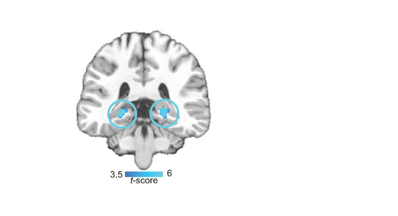
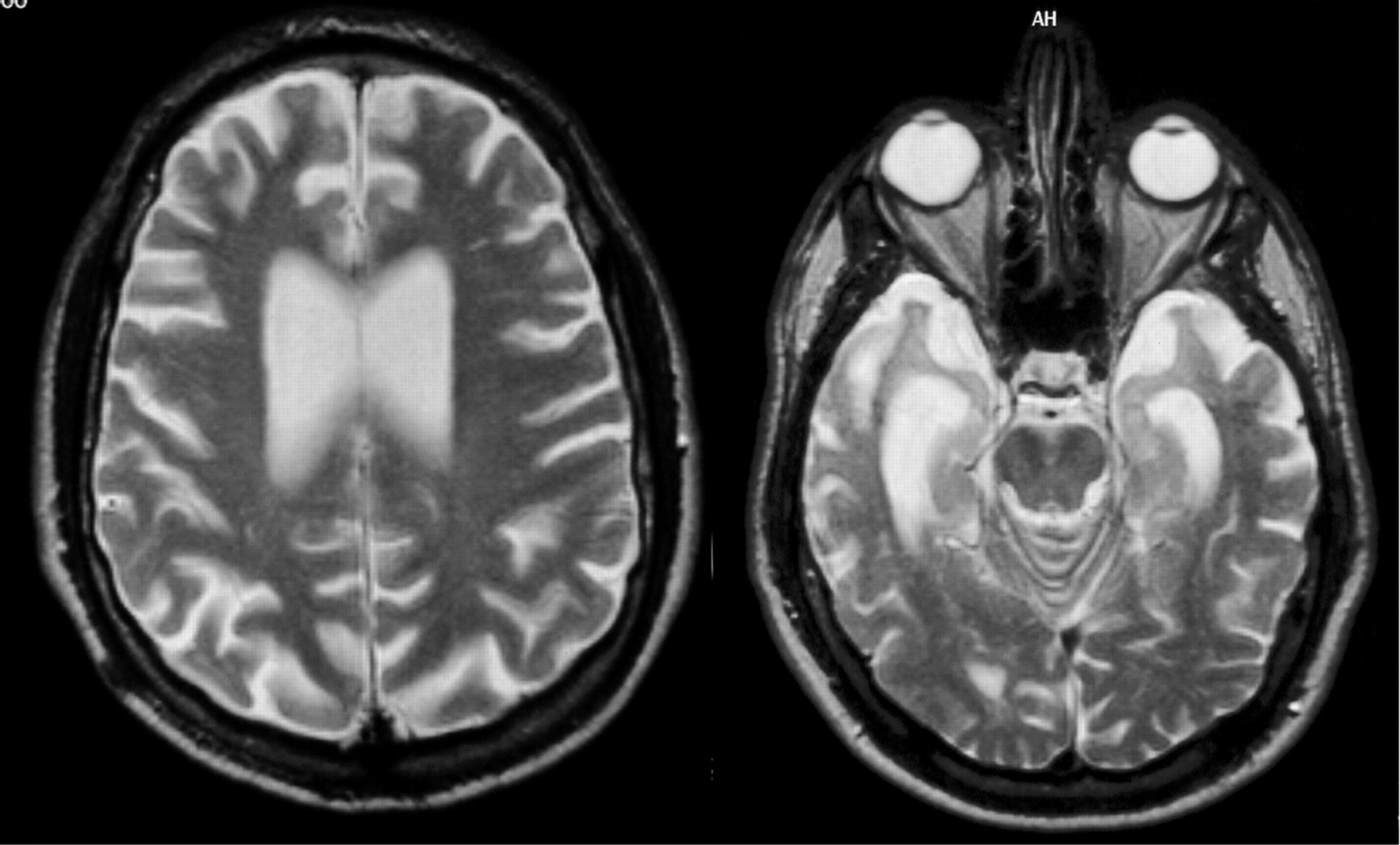
Currently, the definite diagnosis of DAT can only be made through autopsy to find the pathological hallmarks of the disease, microscopic amyloid plaques and neurofibrillary tangles. The development of biomarkers that can reliably indicate presence of the disease at the earliest possible stage is therefore an important public health goal. Macroscopically, DAT is associated with progressive brain tissue loss (Braak and Braak, 1998), which MRI can non-invasively visualise to some extent in-vivo (Thompson et al., 2007). Unsurprisingly, MRI has attracted considerable interest as a tool to identify DAT biomarkers.

Histological studies have shown that the hippocampus is particularly vulnerable to DAT pathology and already considerably damaged at the time clinical symptoms first appear (Braak and Braak, 1998).

Spatial cognition

The “cognitive map theory” proposes that the hippocampus of rats and other animals represents their environments, locations within those environments, and their contents, thus providing the basis for spatial memory and flexible navigation. When it comes to humans, the theory suggests a broader function for the hippocampus, based at least in part on lateralisation of function (Burgess, Maguire and O’Keefe, 2002). The cognitive map theory posits that the hippocampus specifically supports allocentric processing of space in contrast to other brain regions, such as the parietal neocortex, which support egocentric processing (O’Keefe and Nadel, 1978).

Structural MRI scans of the brains of humans with extensive navigation experience, licensed London taxi drivers, were analysed and compared with those of control subjects who did not drive taxis. The posterior hippocampi of taxi drivers were significantly larger relative to those of control subjects. A more anterior hippocampal region was larger in control subjects than in taxi drivers. Hippocampal volume correlated with the amount of time spent as a taxi driver (positively in the posterior and negatively in the anterior hippocampus). These data are in accordance with the idea that the posterior hippocampus stores a spatial representation of the environment and can expand regionally to accommodate elaboration of this representation in people with a high dependence on navigational skills. It seems that there is a capacity for local plastic change in the structure of the healthy adult human brain in response to environmental demands.

Wayfinding

Problems in navigation could even be a good way to diagnose early dementia of Alzheimer type (“DAT”), in future. Virtual reality (“VR”) allows naturalistic evaluation of spatial cognition disorders associated with DAT. These measures seem to be well correlated to daily difficulties of people, thus providing specific measures of cognitive deficits and their functional impact. Thus, VR would be a relevant tool for the early screening of dementia and the differential diagnosis of DAT (Déjos et al., 2011).

While there is abundant evidence for spatial learning and memory decrements in patients with unilateral hippocampal lesions, remarkably little research has been done on spatial memory and learning in patients with DAT, in which relatively selective bilateral hippocampal atrophy is consistently reported in the early stages of the disease (de Pol, 2006). Only a few studies have examined static object-location memory tasks in DAT patients, demonstrating impaired performance compared to controls (Bucks and Willison, 1997; Kessels et al., 2010). Using a real-world wayfinding test, Monacelli and colleagues (Monacelli et al., 2003) investigated a group of DAT patients and demonstrated impaired spatial navigation and spatial orientation in the DAT group, possibly due to an underlying deficit in linking landmark information to route knowledge. Similar findings have also been reported using virtual maze-learning paradigms in AD patients (Cushman, Stein and Duffy, 2008; Kalova et al., 2005).

Current pedestrian navigation systems predominantly use distance-to-turn information and directional information to enable a user to navigate. However, Cherrier and colleagues (Cherrier, Mendez and Perryman, 2001) showed that dementia patients performed better on recognition of landmarks compared with recognition and recall of spatial layout.  Furthermore, relatively few studies have examined the workplaces of staff compared to those that address outcomes for patients and their families. One theme that has been receiving increasing attention over the last few years in the literature about healing environments is wayfinding.

In addition to a complex floor plan, there are other elements that contribute to poor wayfinding and inadequate or conflicting cues such as colours and lighting (Brown, Wright and Brown, 1997). In addition to these elements, clear and understandable wayfinding and maps are fundamental to becoming oriented. However, maps should be oriented so that the top signifies the direction of movement for ease of use (Ulrich et al., 1994). Moreover, the number of signs available has a significant effect on wayfinding along many different measures including travel time, the frequencies of hesitations, the number of times directions were asked, and the reported level of stress. These results suggest that directional signs should be placed at or before every major intersection, at major destinations, and where a single environmental cue or a series of such cues (for instance, a change in flooring material) conveys the message that the individual is moving from one area into another. If there are no key decision points along a route, signs should be placed approximately every 4.6-7.6 m (Ulrich et al., 1994).

Earlier studies reviewed by Day and Calkins (2002) found that much of the orientation work revolved around “signage”, and indentified that personalised and/or unique signage assisted residents in locating desired destinations. Passini and colleagues (Passini et al., 2000) studied newly admitted residents with dementia, and noted that learning new routes was a slow process. Residents who could not identify paths to desired locations exhibited anxiety, confusion, mutism and even panic. They also noted that some residents perceived patterns on the floor as a barrier. They conclude that “capacity of decision-making is reduced to decisions based on immediate and visually accessible information” whether that information was signs, landmarks, or direct visibility of the desired location. They also noted that the typical location of signs is often not seen by residents whose visual field is low to the ground.

Rule, Milke and Dobbs (1991) also found that features such as many similar doorways along corridors, lack of windows to the outside and signage resulted in poorer orientation. McGilton, Rivera and Dawson (2003) conducted a randomised control trial to ascertain the effects of using a locational map and training techniques on the ability of residents to locate distance locations (a dining room on a different floor). While residents in the treatment group showed significant effect within one week of starting the trial, the effect was not sustained three months later.

Driving and DAT

Safe automobile driving requires a driver to perform multiple competing tasks and attend to a host of objects and ongoing events, while simultaneously monitoring traffic with central and peripheral vision to avoid roadway hazards. Impairments of visual acuity and visual fields increase crashes and traffic violations (Burg, 1971). However, drivers with certain neurological conditions may potentially fail to perceive critical roadside targets and dangers even in the absence of a measurable field defect on standard perimetry or diminished visual acuity (Owsley and McGwin, 1999).

Former Urbanites Find Jersey Driving Intimidating

DAT affects processing of visual sensory cues and may produce attentional decline and agnosia (for a review, see Hodges, 2011). These deficits can impair drivers’ processing of visual information such as roadway landmarks and traffic signs that provide key information about a driver’s route, upcoming road hazards, and safety regulations. Uc and colleagues (Uc et al., 2005) studied 33 drivers with probable DAT of mild severity and 137 neurologically normal older adults using a battery of visual and cognitive tests and were asked to report detection of specific landmarks and traffic signs along a segment of an experimental drive. The drivers with mild DAT identified significantly fewer landmarks and traffic signs and made more at-fault safety errors during the task than control subjects.

“The social animal”

“The Social Animal: The Hidden Sources of Love, Character, and Achievement” is a highly celebrated non-fiction book by American journalist David Brooks (Brooks, 2012), who is otherwise best known for his career with The New York Times. The book discusses what drives individual behaviour and decision-making.  Brooks asserts that people’s subconscious minds largely determine who they are and how they behave. He argues that deep internal emotions, the “mental sensations that happen to us”, establish the outward mindset that makes decisions such as career choices. Brooks describes the human brain as dependent on what he calls “scouts” running through a deeply complex neuronal network.

Ultimately, Brooks depicts human beings as driven by the universal feelings of loneliness and the need to belong—what he labels “the urge to merge.” He describes people going through “the loneliness loop” of internal isolation, engagement, and then isolation again. He states that people feel the continual need to be understood by others.

We are, above all, “social animals”, and this is of fundamental importance for wellbeing. For example, Prof. Mario Mendez and Prof. Facundo Manes write recently (Mendez and Manes, 2011), and the authors reviewing this important recent collection of papers on social cognition discuss social cognition dysfunction in a number of different clinical situations, and their potential to give rise to problems in social interactions, immoral or even corrupt behaviour.

Response to stress and resilience

“Resilience” refers to a person’s ability to adapt successfully to acute stress, trauma or more chronic forms of adversity. A resilient individual has thus been tested by adversity (Rutter, 2006) and continues to demonstrate adaptive psychological and physiological stress responses, or `psychobiological allostasis’ (McEwen, 2003; Charney, 2004).

The study of resilience, or stress-resistance, originated in the 1970s with a group of researchers who directed their attention to the investigation of children capable of progressing through normal development despite exposure to significant adversity (Masten, 2001). For many years, research focused on identifying the psychosocial determinants of stress resistance, such as positive emotions, the capacity for self-regulation, social competence with peers and a close bond with a primary caregiver, among other factors (Masten, 1998; Rutter, 1985).

The importance of resilience in policy in living well in dementia, and will be considered further in the final chapter, chapter 18.

Contextual learning

Context-dependence effects are pervasive in everyday cognition. When we perceive objects and colours, we always perceive these among other objects and colours. We listen and speak within other word streams, and every atom of meaning emerges from a background of meanings. Acting appropriately in social interactions requires the interpretation of explicit and implicit contextual clues that orient our responses toward being polite, to make a joke or point out an irony, to say or not say something. Cognitive science and neuroscience research have evidenced context-dependence effects in similar domains of visual perception, emotion, language,  and social cognition in both normal and neuropsychiatric conditions.

Context is important, as shown by the Ebbinghaus illusion which depicts two identical central circles, surrounded by rings of circles. Despite the fact that they are the same size, one circle is perceived as small and the other as big. The contextual information available (the surrounding circles) creates the perception that the center circles are different sizes. This is shown below.

Contextual effects are present at every level, from basic perception to social interaction. This means that we do not perceive objects or process cognitive events in an abstract and universal way. The specific significance of an object, emotion, word, or social situation depends on the contextual effects. During normal cognition, our brains do not process targets and contexts separately; rather, targets are in context.

circles

Behavioural variant frontotemporal dementia and the social context

The “behavioral variant of frontotemporal dementia“ (bvFTD) is characterised by insidiously progressive changes in personality and social interaction that typically precede other cognitive deficits.  Patients may present with compulsiveness, perseverations, or stereotyped repetitive acts, loss of self-consciousness, diminished interest for activities or hobbies, or withdrawal and apathy.  Increased appetite with a tendency for sweet foods is common, and hypersexuality and hyperorality may develop, especially in the advanced stages of the disease.

Early diagnosis is difficult because behavioural problems, invariably reported by friends or family, dominate the clinical picture while cognitive functions are still relatively intact. This is why it is so important to appreciate that dementia does not equal memory problems in every single case (and this is discussed in chapter 18). People with bvFTD often score normally on the Mini-Mental State Examination (“MMSE”), and conventional structural brain imaging (CT and MRI) may not be sensitive to the early changes associated with bvFTD at all. Therefore, early diagnosis relies on clinical interviews and caregiver reports; it can be considerably difficult to distinguish bvFTD from primary psychiatric syndromes.

Patients with bvFTD are now reported consistently to demonstrate reliably deficits in several domains of social cognition such as recognising emotions in facial expressions, empathy processing, decision-making, figurative language, theory of mind, and interpersonal norms.  Little was known about the brains of such patients from an neuroimaging perspective. In particular, given the nature of the cognitive deficits demonstrated by these patients, the authors postulated that, relatively early in the course of the disease, the ventromedial (VMPFC) (or orbitofrontal) cortex is a major locus of dysfunction and that this may relate to the behavioural presentation of these patients clinically described in the individual case histories. A greater definition of the rôle of the ventral frontal cortex, especially given findings in the animal literature, in reversal learning and decision has been a highly influential tranche of research subsequently (Clark, Cools and Robbins, 2004).

At approximately the same time, Lough, Gregory and Hodges (2001) demonstrated relatively intact general neuropsychological and executive function, but extremely poor performance on tasks of theory of mind (ToM). This indicates a dissociation of social cognition and executive function suggesting that in psychiatric presentations of bv-FTD there may be a fundamental deficit in theory of mind independent of the level of executive function. The implications of this finding for diagnostic procedures and possible behavioural management are discussed.

Liu and colleagues (Liu et al., 2004) later compared the behavioral features and to investigate the neuroanatomical correlates of behavioral dysfunction in anatomically defined temporal and behavioural variants of frontotemporal dementia (tvFTD and bvFTD). Volumetric measurements of the frontal, anterior temporal, ventromedial frontal cortical (VMFC), and amygdala regions were made in 51 patients with FTD and 20 normal control subjects, as well as 22 patients with dementia of Alzheimer type (DAT) who were used as dementia controls. FTD patients were classified as bvFTD or tvFTD based on the relative degree of frontal and anterior temporal volume loss compared with controls. Behavioural symptoms, cerebral volumes, and the relationship between them were examined across groups. Both variants of FTD showed significant increases in rates of elation, disinhibition, and aberrant motor behavior compared with DAT. The bvFTD group also showed more anxiety, apathy, and eating disorders, and tvFTD showed a higher prevalence of sleep disturbances than DAT. The only behaviours that differed significantly between bvFTD and tvFTD were apathy, greater in bvFTD, and sleep disorders, more frequent in tvFTD. BvFTD was associated with greater frontal atrophy and tvFTD was associated with more temporal and amygdala atrophy compared with AD, but both groups showed significant atrophy in the VMFC compared with DAT, which was not associated with VMFC atrophy. In FTD, the presence of many of the behavioral disorders was associated with decreased volume in right-hemispheric regions.

Using magnetic resonance imaging (MRI), tensor-based morphometry (TBM), Lu et al. (Lu et al., 2013) was finally used to determine distinct patterns of atrophy between these three clinical groups. The authors concluded that The bvFTD, SV-PPA, and NF-PPA groups displayed distinct patterns of progressive atrophy over a one-year period that correspond well to the behavioral disturbances characteristic of the clinical syndromes. More specifically, the bvFTD group showed significant white matter contraction and presence of behavioral symptoms at baseline predicted significant volume loss of the ventromedial prefrontal cortex. These areas of structural atrophy seem also to be correlated to functional deficits in the case of bvFTD, and now seem to suggest a dissociation in dysfunction even between reversal learning and decision learning deficits at a finer level.

Finally, to complete things, Bertoux and colleagues (Bertoux et al., 2012b) reported that gray matter volume within BA 9 in the medial prefrontal was correlated with scores on the emotion recognition subtest of the he social cognition and emotional assessment”, and the severity of apathetic symptoms in the apathy scale covaried with gray matter volume in the lateral prefrontal cortex (BA 44/45).

The “social context network model”

At a phenomenological level, context-based predictions make social cognition more efficient. Prototypical situations in the environment are represented in “context frames” that integrate information about the meanings of social targets (e.g., an emotional face, a speech) that are likely to appear in a specific scene with information about their relationships.

Ibañez and Manes (2012) proposed that there exists a cortical network that mediates the processing of such contextual associations. This social context network involves regions of the frontal, insular, and temporal cortices. They postulate that frontal areas (e.g., orbitofrontal cortex, lateral prefrontal cortex, superior orbital sulcus) update and associate ongoing contextual information in relation to episodic memory and target-context associations. The temporal regions (amygdala, hippocampus, perirhinal and para-hippocampal cortices) index the value learning of target-context associations. Finally, the insular cortex coordinates internal and external milieus in an internal motivational state. In this way, the insula would provide information integration from internal states and social contexts to produce a global feeling state.

The initial symptoms of FTD reflect the involvement of orbitofrontal cortex as well as the disruption of the rostral limbic system including the insula, the anterior cingulate cortex, the striatum, the amygdala, and the medial frontal lobes. This system is involved in a number of processes such as the evaluation of the motivational or emotional content of internal and external stimuli, error detection, response selection and decision-making, and subsequent regulation of context-dependent behaviours. Recent neuroimaging studies suggest that patients with FTD show predominantly right frontal, anterior insular, and anterior cingulate deterioration, with pronounced orbitofrontal cortex atrophy. Additionally, some studies have reported correlations between behavioural symptoms and brain structures, suggesting that the right orbitofrontal cortex regulates behavior together with a predominantly right-side network involving the insula and striatum. In addition, voxel-based morphometry studies have shown that patients with bvFTD have significant gray matter loss in the anterior insula and in a variety of prefrontal areas.

REFERENCES

Bertoux, M., Volle, E., Funkiewiez, A., de Souza, L.C,, Leclercq, D., and Dubois, B. (2012b) Social Cognition and Emotional Assessment (SEA) is a marker of medial and orbital frontal functions: a voxel-based morphometry study in behavioral variant of frontotemporal degeneration. J Int Neuropsychol Soc, 18(6), pp. 972-85.

Braak H, and Braak E. (1998) Evolution of neuronal changes in the course of Alzheimer’s disease, J Neural Transm Suppl, 53, pp. 127–40.

Brooks, D. (2012) The Social Animal: The Hidden Sources of Love, Character, and Achievement.  London: Random House Trade.

Brown, B., Wright, H., and Brown, C. (1997) A post-occupancy evaluation of wayfinding in 
a pediatric hospital: research findings and implications for instruction,
J Architect Plan Res, 14(1):35e51.

Bucks, R.S., and Willison, J.R. (1997) Development and validation of the Location Learning Test (LLT): a test of visuo-spatial learning designed for use with older 
adults and dementia, Clin Neuropsychol, 11, pp. 273–286.

Burg, A. (1971) Vision and driving: a report on research, Hum Factors, 13, pp. 79–87.

Burgess, N., Maguire, E.A., and O’Keefe, J. (2002) The Human Hippocampus and Review Spatial and Episodic Memory, Neuron, 35, pp. 625–641.

Charney, D.S. (2004) Psychobiological mechanisms of resilience and vulnerability: implications for successful adaptation to extreme stress, Am. J. Psychiatry, 161, pp. 195–216.

Clark, L., Cools, R., and Robbins, T.W. (2004) The neuropsychology of the ventral prefrontal cortex: decision-making and reversal learning, Brain and Cognition, 55, 41-53.

Cherrier, M., Mendez, M., and Perryman, K. (2001) Route Learning Performance in Alzheimer Disease Patients, Neuropsychiatry, Neuropsychology, and Behavioral Neurology, 14:3, pp. 159– 168.

Cushman, L.A., Stein, K., and Duffy C.J. (2008) Detecting navigational deficits in cognitive aging and Alzheimer disease using virtual reality, Neurology, 71, pp. 888–895.

Day, K and Calkins, MP. (2002) Design and Dementia, in: Churchman, R.B.A. (ed.) Handbook of Environmental Psychology, New York: John Wiley & Sons.

de Pol, L.A., van der Flier, W.M., Korf, E.S., Fox, N.C., Barkhof, F., and Scheltens, P. (2007) Baseline predictors of rates of hippocampal atrophy in mild cognitive impairment, Neurology, 69, pp. 1491–7.

Déjos, M., Sauzéon, H., Falière, A., and N’Kaoua, B. (2011) Naturalistic assessment of spatial cognition disorders in Alzheimer’s disease using virtual reality: Abstracts [CO37-005–EN], Annals of Physical and Rehabilitation Medicine, 54S, e87–e94 (p. e90).

Ferri, C.P., Prince, M., Brayne C, Brodaty, H., Fratiglioni, L., Ganguli, M., Hall, K., Hasegawa, K., Hendrie, H., Huang, Y., Jorm, A., Mathers, C., Menezes, P.R., Rimmer, E., and Scazufca, M; Alzheimer’s Disease International. (2005) Global prevalence of dementia: a Delphi consensus study, Lancet, 366, pp. 2112–7.

Hodges, J.R. (2010) Chapter 24.2.2 Dementia, in London, in Warrell, D.A., Cox, T.M., and Firth, J.D. (eds.) Oxford Textbook of Medicine, Oxford, UK (5th edition): Oxford University Press.

Ibañez, A. and Manes, F. (2012) Views and reviews: Contextual social cognition and the behavioral variant of frontotemporal dementia, Neurology, 78, pp. 1354–1362.

Kalová, E., Vlcek, K., Jarolímová, E., and Bures, J. (2005) Allothetic orientation and sequential ordering of places is impaired in early stages of Alzheimer’s disease: corresponding results in real space tests and computer tests, Behav Brain Res, 159: 175–186.

Kessels, R.P.C., Rijken, S., Joosten-Weyn Banningh, L.W.A., Van Es, N., and Olde Rikkert, M.G.M. (2010) Categorical spatial memory in patients with Mild Cognitive Impairment and Alzheimer dementia: positional versus object-location recall, J Int Neuropsychol Soc, 16, pp. 200–204.

Liu, W., Miller, B.L., Kramer, J.H., Rankin, K., Wyss-Coray, C., Gearhart, R., Phengrasamy, L., Weiner, M., and Rosen, H.J. (2004) Behavioral disorders in the frontal and temporal variants of frontotemporal dementia, Neurology, 62(5), pp. 742-8.

Lough, S., Gregory, C., and Hodges, J.R. (2001) Dissociation of social cognition and executive function in frontal variant frontotemporal dementia, Neurocase, 7(2), pp. 123-30.

Lu, P.H., Mendez, M.F., Lee, G.J., Leow, A.D., Lee, H.W., Shapira, J., Jimenez, E., Boeve, B.B., Caselli, R.J., Graff-Radford, N.R., Jack, C.R., Kramer, J.H., Miller, B.L., Bartzokis, G., Thompson, P.M., and Knopman, D.S. (2013) Patterns of brain atrophy in clinical variants of frontotemporal lobar degeneration, Dement Geriatr Cogn Disord, 35(1-2): pp. 34-50.

Maguire, E.A., Gadian, D.G., Joshnsrude, I.S., Good, C.D., Ashburner, J., Frackowiak, R.S.J., and Frith, C.D. (2000) Navigation-related structural change in the hippocampi of taxi drivers, Procl Acad Nat Sci, 97(8), pp. 4398-403.

Masten, A.S. (2001) Ordinary magic. Resilience processes in development. Am Psychol, 56, pp. 227–238.

Masten, A.S., and Coatsworth, J.D. (1998) The development of competence in favorable and unfavorable environments. Lessons from research on successful children, Am Psychol, 53, pp. 205–220.

McEwen, B.S. (2003) Mood disorders and allostatic load, Biol Psychiatry, 54, pp. 200–207.

McGilton, K.S., Rivera, T.M., and Dawson,, R. (2003) Can we help persons with dementia find their way in a new environment?, Aging and Mental Health, 7, pp. 363-71.

Mendez, M.F., Manes, F. (2011) The emerging impact of social neuroscience on neuropsychiatry and clinical neuroscience, Soc Neurosci, 6(5-6), pp. 415-9.

Monacelli, A.M., Cushman, L.A., Kavcic, V., and Duffy C.J. (2003) Spatial disorientation in Alzheimer’s disease: the remembrance of things passed, Neurology, 61, pp. 1491–1497.

O’Keefe, J., and Nadel, L. (1978) The hippocampus as a cognitive map. Oxford: Oxford University Press.

Owsley, C., and McGwin, G. (1999) Vision impairment and driving, Surv Ophthalmol, 43, pp. 535–50.

Passini, R., Pigot, H., Rainville, C., Tétreault, M.H. (2000) Wayfinding in a Nursing Home for Advanced Dementia of the Alzheimer’s Type, Environment & Behavior, 32, pp. 684–710.

Rule, B.G., Milke, D.L., Dobbs, A.R. (1992) Design of institutions: Cognitive functioning and social interactions of the aged resident, Journal of Applied Gerontology, 11, pp. 475–488.

Rutter, M. (1985) Resilience in the face of adversity: protective factors and resistance to psychiatric disorder, Br J Psychiatry, 147, pp. 598–611.

Rutter, M. (2006) Implications of resilience concepts for scientific understanding, Ann NY Acad Sci, 1094, pp. 1–12.

Thompson, P.M., Hayashi, K.M., Dutton, R.A., Chiang, M.C., Leow, A.D., Sowell, E.R., De Uc, E.Y., Rizzo, M.,  Anderson, S.W., Shi, Q., and Dawson, J.D. (2005) Driver landmark and traffic sign identification in early Alzheimer’s disease, J Neurol Neurosurg Psychiatry, 76, pp. 764–768.

Ulrich, R.S., Quan, X., Zimring, C., Joseph, A., and Choudhary, R. (2004) The role of the physical 
environment in the hospital of 21st century: a once-in-a-lifetime opportunity. Concord: CA: Center for Health Design.

van de Pol, L.A., Hensel, A., van der Flier, W.M., Visser, P.J., Pijnenburg, Y.A., Barkhof, F., Gertz, H.J., and Scheltens, P. (2006) Hippocampal atrophy on MRI in frontotemporal lobar degeneration and Alzheimer’s disease, J Neurol Neurosurg Psychiatry, 77, pp. 439–442.

Zubicaray, G., Becker, J.T., Lopez, O.L., Aizenstein, H.J., and Toga A.W. (2007) Tracking Alzheimer’s Disease, Ann N Y Acad Sci, 1097, pp. 183-214. [Review.]