There’s something about tau

Tau

Alzheimer’s disease is the most prevalent type of dementia globally, and therefore traditionally gets the most focus. However, there are other neurodegenerative conditions of note which are of massive importance. For example, neurodegeneration with brain iron accumulation (NBIA) is a group of disorders characterized by dystonia, parkinsonism and spasticity. Models of Alzheimer’s disease, frontotemporal dementia, Parkinson’s disease and Huntington’s disease show some striking similarities to the corresponding human pathologies in terms of axonal transport disruption, protein aggregation, synapse loss and some behavioural phenotypes (Gilley, Adalbert and Coleman, 2011). In early stages of Alzheimer’s disease, neurofibrillary tangles (NFT) are largely restricted to the entorhinal cortex and medial temporal lobe. At later stages, when clinical symptoms generally occur, NFT involve widespread limbic and association cortices. At this point in the disease, amyloid plaques are also abundantly distributed in the cortex.

Induced pluripotent stem cells (also known as iPS cells or iPSCs) are a type of pluripotent stem cell that can be generated directly from adult cells. The iPSC technology was pioneered by Shinya Yamanaka’s lab in Kyoto, Japan, who showed in 2006 that the introduction of four specific genes encoding transcription factors could convert adult cells into pluripotent stem cells. He was awarded the 2012 Nobel Prize along with Sir John Gurdon “for the discovery that mature cells can be reprogrammed to become pluripotent.”

Pluripotent stem cells hold great promise in the field of regenerative medicine. Because they can propagate indefinitely, as well as give rise to every other cell type in the body (such as neurons, heart, pancreatic, and liver cells), they represent a single source of cells that could be used to replace those lost to damage or disease.

According to Wray and Fox (2016),

“It is the use of stem cells as disease models that perhaps has the most to offer in terms of immediate gain, and the most exciting development is the potential to assay potential therapeutics with induced pluripotent stem cells (iPSCs).”

But Wray and Fox (2016) later comment:

“Of particular relevance to Alzheimer’s disease is the fi ending that the expression profile of tau remains feral-like in iPSC-derived neurons until 1 year in culture. Even in cases of familial disease with the earliest onset, the disease only manifests clinically several decades after the onset of pathology and structural changes—how effectively will iPSCs recapitulate the full time course of disease-associated molecular changes?”

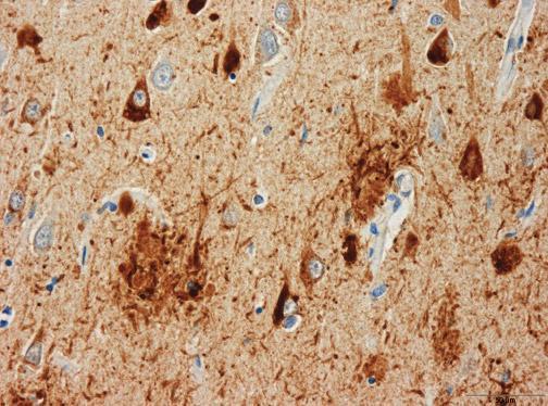
Tau proteins (or τ proteins, after the Greek letter by that name) are proteins that stabilise parts of the cell called “microtubules”. They are abundant in neurons of the central nervous system and are less common elsewhere, but are also expressed at very low levels in CNS astrocytes and oligodendrocytes. Pathologies and dementias of the nervous system such as Alzheimer’s disease are associated with tau proteins that have become defective and no longer stabilise microtubules properly.

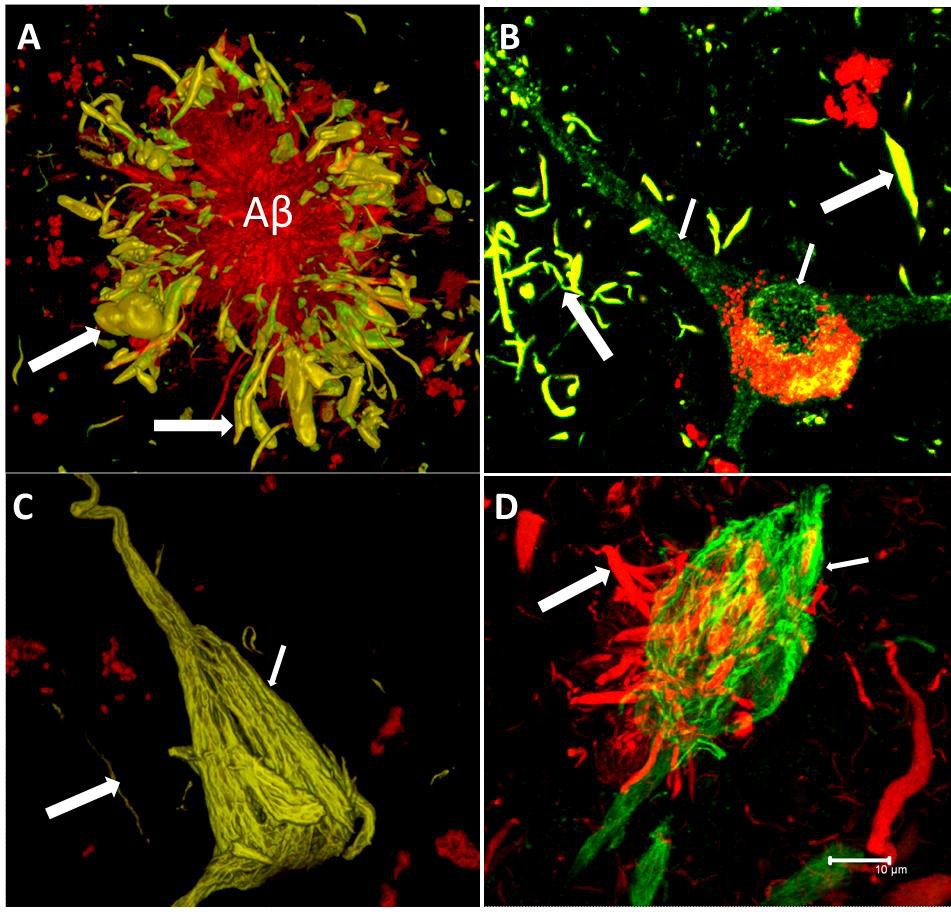
The tau hypothesis of Alzheimer’s disease states that excessive or abnormal phosphorylation of tau results in the transformation of normal adult tau into PHF-tau (paired helical filament) and NFTs (neurofibrillary tangles). But it is clearly more complicated than that. Deposition of highly phosphorylated tau in the brain is the most significant neuropathological and biochemical characteristic of the group of neurodegenerative disorders termed the tauopathies. Pathological hyperphosphorylation of the microtubule-associated protein tau is characteristic of Alzheimer’s disease and the associated tauopathies. The reciprocal relationship between phosphorylation and O-GlcNAc modification of tau and reductions in O-GlcNAc levels on tau in AD brain offers motivation for the generation of potent and selective inhibitors that can effectively enhance O-GlcNAc in vertebrate brain (Yuzwa et al., 2008).

The discovery of tau fragments in these diseases suggests that tau cleavage and tau phosphorylation, both of which induce conformational changes in tau, could each have roles in disease pathogenesis. As Hanger and Wray (2010) note, the identities of the proteases responsible for degrading tau, resulting in the appearance of truncated tau species in physiological and pathological conditions, are not known.

The Bavarian psychiatrist Alois Alzheimer is traditionally credited with the first description of the most characteristic pathological brain change—neurofibrillary tangles (NFT)—of a yet-unnamed disease in a 51-year-old woman from Frankfurt am Main, who had developed dementia.

During the 1990s, the significance of tau pathology for neurodegenerative diseases, in particular for dementia of the Alzheimer Type, remained in the shadow of the amyloid based approaches. However, as the distribution pattern and overall quantity of amyloid turned out to be of limited significance for pathological staging of AD progression and symptom severity, and after detailed studies of the maturation and distribution of NFTs showing correlation with degree of cognitive decline and memory impairment in Alzheimer’s disease, Braak and Braak proposed a neuropathological staging of the gradual deposition of abnormal tau within vulnerable neurons in brain areas in the form of either NFT or neuropil threads. Post-mortem Braak staging of neurofibrillary tau tangle topographical distribution is one of the core neuropathological criteria for the diagnosis of Alzheimer’s disease.

Based on the biochemically diverse range of pathological tau proteins, Šimić and colleagues (2006) have recently reviewed a number of different approaches which have been proposed to develop new potential therapeutics. Here we discuss some of the most promising ones: inhibition of tau phosphorylation, proteolysis and aggregation, promotion of intra- and extracellular tau clearance, and stabilization of microtubules.

The recent development of candidate PET imaging tracers targeting aggregated tau (now enables not only the brain burden but also the anatomical distribution of tau pathology to be mapped directly in living subjects. One such PET tracer, 18F-AV-1451 (also known as 18F-T807), has been shown to bind selectively to paired-helical filament (PHF) tau, and to exhibit favourable kinetics, low non-specific binding and differential uptake in Alzheimer’s disease versus healthy control subjects. It has been very recently been reported that in vivo 18F-AV-1451 positron emission tomography images across the Alzheimer’s disease spectrum can be classified into patterns similar to those prescribed by Braak neuropathological staging of tau pathology (Schwarz et al., 2016).

But there’s more to tau than Alzheimer’s disease. In NBIA, iron accumulates in the basal ganglia and may be accompanied by Lewy bodies, axonal swellings and hyperphosphorylated tau depending on NBIA subtype (Arber et al., 2015). And there’s more to Alzheimer’s disease than tau. For example, results from Pooler and colleagues (Pooler et al., 2015) strongly support the hypothesis that cortical amyloid accelerates the spread of tangles throughout the cortex and amplifies tangle-associated neural system failure in AD. The story is gradually though unravelling.

Talk

Dr Selina Wray will be giving a presentation at 4 pm today in session 11 of the Alzheimer’s Research UK Research Conference entitled “Modelling tauopathy in patient-derived neutrons: good things come to those who wait?” (link here).

Recommended reading

Arber CE, Li A, Houlden H, Wray S. Insights into molecular mechanisms of disease in neurodegeneration with brain iron accumulation: unifying theories. Neuropathol Appl Neurobiol. 2015 Apr 14. doi: 10.1111/nan.12242. [Epub ahead of print].

Gilley J, Adalbert R, Coleman MP. Modelling early responses to neurodegenerative mutations in mice. Biochem Soc Trans. 2011 Aug;39(4):933-8. doi: 10.1042/BST0390933.

Hanger DP, Wray S. Tau cleavage and tau aggregation in neurodegenerative disease. Biochem Soc Trans. 2010 Aug;38(4):1016-20. doi: 10.1042/BST0381016.

Schwarz AJ, Yu P, Miller BB, Shcherbinin S, Dickson J, Navitsky M, Joshi AD, Devous MD Sr, Mintun MS Regional profiles of the candidate tau PET ligand 18F-AV-1451 recapitulate key features of Braak histopathological stages. Brain. 2016 Mar 2. pii: aww023. [Epub ahead of print].

Šimić G, Babić Leko M, Wray S, Harrington C, Delalle I, Jovanov-Milošević N, Bažadona D, Buée L, de Silva R, Di Giovanni G, Wischik C, Hof PR. Tau Protein Hyperphosphorylation and Aggregation in Alzheimer’s Disease and Other Tauopathies, and Possible Neuroprotective Strategies. Biomolecules. 2016 Jan 6;6(1). pii: E6. doi: 10.3390/biom6010006.

Wray S, Fox NC. Stem cell therapy for Alzheimer’s disease: hope or hype? Lancet Neurol. 2015 Dec 15. pii: S1474-4422(15)00382-8. doi: 10.1016/S1474-4422(15)00382-8. [Epub ahead of print].

Yuzwa SA, Macauley MS, Heinonen JE, Shan X, Dennis RJ, He Y, Whitworth GE, Stubbs KA, McEachern EJ, Davies GJ, Vocadlo DJ.29. A potent mechanism-inspired O-GlcNAcase inhibitor that blocks phosphorylation of tau in vivo. Nat Chem Biol. 2008 Aug;4(8):483-90. doi: 10.1038/nchembio.96. Epub 2008 Jun.

“Amyloid busting drugs” – true weapons of mass destruction, or blatant puffery?

I must admit I haven’t got anything to gain from the ‘amyloid hypothesis of Alzheimer’s disease’.

I am not a paid researcher to do with dementia, nor am I paid to promote research campaigns. I do not work for any dementia charities.

In fact, you are entitled to think I am pretty useless. But I am not in fact. I care passionately about dementia research, having done my own work in discovering an innovative way to diagnose the behavioural variant of frontotemporal dementia at Cambridge between 1997-2000. In fact, a key supervisor of my Ph.D. was Prof John Hodges who won a lifetime achievement award at this year’s Alzheimer’s Association conference held in Washington. I consider Hodges a friend as well as colleague. I have been open with him about my personal and professional background. Indeed, John, still knowing all the facts, did me the honour of writing the main Foreword to my first book, “Living well with dementia: the importance of the person and the environment.”


So what is the ‘amyloid hypothesis of Alzheimer’s disease‘?  This is, in a nutshell, a build up of an abnormal protein in the brain, as plaques. This build up is like mini brillo pads clogging up the brain, causing the brain to shrink, and buggering up its overall function.

That was my best at being a paid scientific communicator.

But this hypothesis is not without its problems. One key problem is the seminal finding of Price and colleagues (2009) that up to 40% of people without dementia can reach ‘neuropathological criteria’ for Alzheimer’s disease.

And there are in fact perfectly sensible explanations why the amyloid plaques which build up are not the cause of the problems in themselves.


These are summarised well in Morris, Clark and Vissel (2014).  It might be that it is type of amyloid plaque which is important to decline; some plaques are non-toxic; amyloid plaques are not actually the cause of Alzheimer’s disease (but soluble beta amyloid might be); and so on.

The amyloid treatments, or else “anti-Aβ treatments’, may have in fact interesting reasons why the “drugs don’t work”.

Back to Morris, Clark and Vissel (2014).

“So far, anti-Aβ treatments have broadly failed to meet their primary clinical endpoints and some major phase 3 trials were halted early. None of the tested treatments have produced a discernible functional recovery, or altered the course of disease. In fact alarmingly some, specifically inhibitors of γ-secretase, lead to an increased decline in cognition.

Another prominent suggested reason for clinical failures of anti-Aβ drugs in particular are that the agents used initially were not properly validated and were flawed. A recent study has shown the monoclonal anti-Aβ antibodies, solanezumab and crenezumab, fail to target human Aβ as effectively as they target over-expressed human Aβ in mouse models. The possibility was also countenanced that only amyloid plaques, potentially functionally inert, rather than soluble Aβ oligomers were targeted in early trials. Furthermore monotherapies may not be capable of effectively reducing Aβ plaque load. A double pronged approach to reduce Aβ by both active immunisation and inhibition of β-secretase has effectively cleared plaques in mice. However, as reviewed recently, therapeutic approaches targeting plaque and approaches targeting soluble Aβ have both now been tested in humans, with equally negative outcomes.”


One idea which had become in vogue was that it was the artist known as ‘fibrillar β-amyloid (Aβ)’  which was the culprit in Alzheimer’s disease. Look at what Stonnington and colleagues reviewed in 2014.

Fibrillar β-amyloid (Aβ) imaging, most notably with [11C]-benzothiazole radiotracer Pittsburgh compound B (PiB) PET and more recently with the fluorine-18-labeled tracers such as Florbetapir, has emerged as a potential biomarker for preclinical AD. Evidence suggests that increases in fibrillar Aβ deposition precede neuronal injury, and fibrillar amyloid deposition is a potential predictor of later symptomatic cognitive decline. We now have evidence that preclinical cognitive decline correlates with an increased measure of fibrillar Aβ deposition and that this effect is independent of APOE status.


So we have a bit of a problem on our hands. International scientists, and ‘interested parties’, convening in Washington, for the Alzheimer’s Association International Conference 2015, even if battled were on the whole extremely optimistic. I am not going to call it a trade fair. It is meant to be, a rather, serious discussion of the results, as well as methodological concerns, of rather expensive experiments in the “rolls-royce” end of dementia.

It’s well known that the relationship between the media and reporting of dementia has been in the past a tricky one. Take, for example, this response from the Association of Medical Research Charities, Cancer Research UK and the Wellcome Trust to the ‘Leveson inquiry: Culture, practice and ethics of the press’, dated January 2012.

Hype and false hope: The flipside of the health scare is the overcooked breakthrough. Many newspapers (though not all of them) are apt to exaggerate interesting but preliminary advances in biomedical science, proclaiming them as groundbreaking achievements that will transform individuals’ health when in fact they are reporting nothing more than promising results from experiments on mice, or cells grown in culture. 


 Such reporting can have several negative consequences. First, it raises expectations for advances in medical science, many of which will fall by the wayside over the long journey from laboratory bench to patient bedside. This can feed a public perception that science is always promising and never delivering. 


Secondly, and more worryingly, it can often raise false hope among patients. This is particularly true and damaging where it concerns treatments for incurable diseases that are not proven, yet which are portrayed as “miracle cures”. This can lead patients to spend life savings on treatments that are most unlikely to work, or on occasion to eschew the most effective known therapies in favour of alternatives that are untested or disproved.

So we were faced with a difficult situation earlier this week with solanezumab (Lilly).


Key paragraphs from the press release this week are given thus.

Showing that an investigational treatment has slowed the progression of a degenerative brain disease like Alzheimer’s is extremely challenging. Researchers have proposed overcoming this problem with a type of study called a “delayed-start” trial. In delayed-start studies, patients are randomly assigned to start active treatment at the beginning of the study or are placed in a “delayed-start” group that receives a placebo treatment for a period of time before being given the active experimental therapy. Researchers then compare the two groups at a later, pre-defined point in time to assess their response to the treatment.


If the treatment can actually slow disease progression, both groups will benefit, but the group that started active treatment later in the study will have progressed further in the disease before they got the drug – while they were on placebo. As a result, the late starters will not be able to “catch up” to the group whose disease progression was slowed for the full duration of the study.


Treatment differences at 28 weeks in EXP-EXT between the early start and delayed start groups for cognition (ADAS-Cog14) and function (ADCS-iADL) were similar to differences at the end of the placebo-controlled period, within a pre-defined margin. In other words, the delayed starters did not “catch up.

And why 28 weeks?

The answer is actually blindingly obvious – this should be the correct length of time for the drug to have some effect, and our understanding of this is totally dependent on our knowledge of how this drug is metabolised.

Let’s assume that this figure is correct. But even this is not clear-cut.

Turn for a moment to a paper from Liu-Sefert and colleagues this year,

In a delayed-start design, the placebo-controlled period should be sufficiently long to allow a disease-modifying drug to show an effect and the delayed-start period should be long enough to observe a symptomatic effect. Most clinical studies testing potential disease-modifying treatments have used 18 months for the placebo-controlled period, as in the EXPEDITION studies. Given that the half-life of solanezumab is approximately 28 days, a duration of 28 weeks (approximately 6 months) was chosen for the delayed-start period for the primary efficacy analyses of EXPEDITION-EXT to ensure that the duration of the period was over 5 half-lives and was thus adequate for delayed-start patients to achieve pharmacokinetic equilibrium. The literature also suggests that most symptomatic drugs reach peak effect in less than 6 months. EXPEDITION-EXT is still ongoing and future analyses will demonstrate whether the effect persists beyond 28 weeks.”


Do you remember the fibrillar amyloid which might have an effect?

Well, read for a moment a description of solanezumab thus.

Solanezumab is a humanized monoclonal IgG1 antibody directed against the mid-domain of the Aβ peptide. It recognizes soluble monomeric, not fibrillar, Aβ. The therapeutic rationale is that it may exert benefit by sequestering Aβ, shifting equilibria between different species of Aβ, and removing small soluble species of Aβ that are directly toxic to synaptic function. In preclinical research, a single injection of m266, the mouse version of solanezumab, reversed memory deficits in APP-transgenic mouse models while leaving amyloid plaques in place, raising the prospect of targeting the soluble pool of Aβ.


So it doesn’t recognise fibrillar Aβ, but it can reverse memory deficits?

The take-home message there, previously reported only last year, was not good from the phase 3 clinical trial, from Doody and colleagues (Doody et al., 2014):

Two randomized, double-blind, placebo-controlled, phase 3 studies of solanezumab treatment were performed in patients with mild-to-moderate Alzheimer’s disease. … Solanezumab, a humanized monoclonal antibody that binds amyloid, failed to improve cognition or functional ability.


Undergraduate students and higher are told that the fundamental problem in Alzheimer’s disease is in the hippocampus, or at least the parahippocampal areas, as this is the heart of learning and memory systems. And yet in the original study the following observation was made (Doody et al., 2014):

However, the current studies failed to show treatment effects on hippocampal or total brain volumes or on amyloid accumulation with the use of 18 F-florbetapir–PET. These results are consistent with the observation that solanezumab does not target fibrillar amyloid. Our PET findings were not conclusive because of the small sample, but sufficient numbers of solanezumab-treated and untreated patients underwent serial MRI to make the failure to detect a slowing of brain atrophy a meaningful finding.

The inclusion criteria for the actual trial though is interesting (Doody et al., 2014), and this presumably are the inclusion criteria for the follow up presented at #AAIC2015 this week.

Both trials involved otherwise healthy patients 55 years of age or older who had mild-to-moderate Alzheimer’s disease without depression. Mild-to-moderate Alzheimer’s disease was documented on the basis of a score of 16 to 26 on the Mini–Mental State Examination (MMSE; score range, 0 to 30, with higher scores indicating better cognitive function)  and the criteria of the National Institute of Neurological and Communicative Disorders and Stroke–Alzheimer’s Disease and Related Disorders Association.”

and depression was excluded.

How many clinical raters? If there were more than 1, did they show good agreement? How accurate is a combined score of the MMSE and NINCDS-ADRDA criteria in producing a definitive diagnosis of Alzheimer’s disease? There will be some – and we don’t know how many – who will not have a diagnosis of Alzheimer’s at all in the Alzheimer’s disease randomised patients group.

Vellas and colleagues (2013) had previously warned about a biomarker problem in looking at the methodological issues of solanezumab in doing a ‘lessons learned’ type of report:

Solanezumab, another humanized monoclonal antibody, targets an epitope in the middle of the Ab peptide. The predominant message emerging from results of the two phase 3 trials (EXPEDITION 1 and 2) is that targeting amyloid appears to have a positive effect on cognition in mild AD, although there were no biomarker changes that indicated a treatment effect.”


As it happens, if the Strictly judges make or break Strictly contestants, it is a concern that investors were not that impressed with the AAIC2015 outcomes. An underwhelming performance of solanezumab was described here this week. Apparently, Lilly shares fell 3.3 percent in early trading.

All of this smacks somewhat of puffery.

I asked somebody who has quite a strong family history of dementia, and a Daily Express reader, what she thought of a possible research development in a cure. She answered straight away, “I think it’s brilliant. Who wouldn’t want a cure for dementia?”

And I have a lot of sympathy with this view. But in law, puffery is a promotional statement which presents subjective rather than objective views. The defense of the media, even reputable broadcasters, that in the relative absence of ‘good news’ about dementia, ignoring completely the vast field of living better with dementia that I and many work in, there is a need to grasp onto any titbits of good news.

But hyperbole is ‘exempt’ from the normal standards of relying on a statement to make a decision.

This is established in English law in a seminal case (albeit only from the Court of Appeal), but the same principles currently apply in other jurisdictions, in the late 19th Century (Carlill v Carbolic Smoke Ball Company 1892).

The United States Federal Trade Commission (FTC) defined puffery as a “term frequently used to denote the exaggerations reasonably to be expected of a seller as to the degree of quality of his product, the truth or falsity of which cannot be precisely determined.”

A “puff piece” is an idiom for “a journalistic form of puffery: an article or story of exaggerating praise that often ignores or downplays opposing viewpoints or evidence to the contrary“.

But there are 47 million people in the world who are relying on news of developments in Alzheimer’s disease and the 100 or so other types of dementia. Some of the Alzheimer’s disease research community have now successfully dug themselves into a hole.


I would strongly urge them to stop digging.


References

Doody RS, Thomas RG, Farlow M, Iwatsubo T, Vellas B, Joffe S, Kieburtz K, Raman R, Sun X, Aisen PS, Siemers E, Liu-Seifert H, Mohs R; Alzheimer’s Disease Cooperative Study Steering Committee; Solanezumab Study Group.Phase 3 trials of solanezumab for mild-to-moderate Alzheimer’s disease. N Engl J Med. 2014 Jan 23;370(4):311-21.

Liu-Seifert H, Andersen SW, Lipkovich I, Holdridge KC, Siemers E (2015) A Novel Approach to Delayed-Start Analyses for Demonstrating Disease- Modifying Effects in Alzheimer’s Disease. PLoS ONE 10(3): e0119632. doi:10.1371/journal.pone.0119632

Morris GP, Clark IA, Vissel B. Inconsistencies and controversies surrounding the amyloid hypothesis of Alzheimer’s disease.  Acta Neuropathol Commun. 2014 Sep 18;2:135. doi: 10.1186/s40478-014-0135-5.

Price JL, McKeel DW Jr, Buckles VD, Roe CM, Xiong C, Grundman M, Hansen LA, Petersen RC, Parisi JE, Dickson DW, Smith CD, Davis DG, Schmitt FA, Markesbery WR, Kaye J, Kurlan R, Hulette C, Kurland BF, Higdon R, Kukull W, Morris JC (2009) Neuropathology of nondemented aging: Presumptive evidence for preclinical Alzheimer disease. Neurobiol Aging 30:1026–1036.

Stonnington CM, Chen K, Lee W, Locke DE, Dueck AC, Liu X, Roontiva A, Fleisher AS, Caselli RJ, Reiman EM. Fibrillar amyloid correlates of preclinical cognitive decline. Alzheimers Dement. 2014 Jan;10(1):e1-8. doi: 10.1016/j.jalz.2013.01.009. Epub 2013 Apr 11.

Vellas B, Carrillo MC, Sampaio C, Brashear HR, Siemers E, Hampel H, Schneider LS, Weiner M, Doody R, Khachaturian Z, Cedarbaum J, Grundman M, Broich K, Giacobini E, Dubois B, Sperling R, Wilcock GK, Fox N, Scheltens P, Touchon J, Hendrix S, Andrieu S, Aisen P; EU/US/CTAD Task Force Members. Designing drug trials for Alzheimer’s disease: what we have learned from the release of the phase III antibody trials: a report from the EU/US/CTAD Task Force. Alzheimers Dement. 2013 Jul;9(4):438-44. doi: 10.1016/j.jalz.2013.03.007.


Puffery discussion acknowledgement to Peter Watt.

Is a new sophisticated brain scan desirable to diagnose dementia?

Amyloid plaques

Wouldn’t it be lovely Prime Minister, David Cameron MP, could announce a breakthrough which nails the problem of the diagnosis of Alzheimer’s disease?

The definitive diagnosis of dementia of the Alzheimer type (DAT) comes post mortem (though in practice various techniques while the patient is alive can be used to tell whether a patient has a type of dementia).

The full armoury of tests includes thinking tests or cognitive neuropsychology, a sample of the fluid from the spine (cerebrospinal fluid), the clinical history and examination of the patient, brain waves (the EEG), or even (rarely) a brain biopsy; that’s even before considering types of scan, like the ‘CT scan’, the ‘MRI scan’, or ‘functional scan’.

The trick of the clinician, varying with levels of expertise, is to make the diagnosis reliably such that a person living with dementia might be able to ‘access’ appropriate care in the system; and those without dementia aren’t given an incorrect label of ‘dementia’.

DAT is one of the hundreds of causes of dementia (although most of the media use ‘Alzheimer’s Disease’ and dementia unhelpfully synonymously.)

Amyloid build-up and the diagnosis

There has been a popular idea that the build up of a substance called amyloid which builds up in the brain might hold the clue to early diagnosis of Alzheimer’s Disease.

In recent years, the emphasis has swung to ‘timely diagnosis’, with the national clinical lead for dementia, Prof Alistair Burns, emphasising that the diagnosis should be made at a time appropriate for the person himself or herself.

A ‘quick fix’ in a test for DAT seems very attractive, but it’s important to remember that the dementia of the Alzheimer type is only one (but the most common) cause of dementia across all age groups.

How to use the test in a safe way

The way in which this diagnosis could be made has also come under scrutiny.  A method which uses a radioactive label to which at how much label can bind to abnormal amyloid in the brain, to be practical, should not be excessively time-consuming to administer. It also should not be prohibitively expensive.

Also critically, it should be reliable. In other words, it shouldn’t show up ‘positives’ in otherwise well people, who never go onto develop dementia. A critical problem is that there are many causes of memory loss in older people, including of course depression.

To make things even more complicated, there is a very interesting group of people whose thinking and memory are normal, even late in life, yet their brains are full of amyloid beta plaques that appear to be identical to what’s seen in dementia of the Alzheimer type. How this can occur is an important clinical research question.

Hard plaques made of a protein called amyloid beta are always present in the brain of a person diagnosed with the dementia of the Alzheimer type.  But the simple presence of plaques does not always result in impaired thinking and memory. In other words, the plaques are necessary – but not sufficient – to cause DAT.

Is it the type of amyloid which matters?

Earlier this year a paper was published in the prestigious journal in the US (Esparza TJ, Zhao H, Cirrito JR, Cairns NJ, Bateman RJ, Holtzman DM, Brody DL. (2013) Amyloid-β oligomerization in Alzheimer dementia versus high-pathology controls. Ann Neurol. 73(1):104-19. doi: 10.1002/ana.23748. Epub 2012 Dec 7.)

An important clue may come from still come from a form of amyloid beta, but not necessarily in the form of plaques. Instead, smaller molecules of amyloid beta appear more closely correlated with whether a person develops symptoms of dementia; these are called “amyloid beta oligomers“.

Earlier this year, this group developed a way of measuring  these amyloid beta oligomers in minute quantities, without binding to similar things.

These amyloid beta oligomers were detected in samples of brain from patients with DAT and nondemented patients with amyloid plaque pathology. However, amyloid beta oligomer concentrations in samples from patients with DAT were tightly correlated with amyloid plaque coverage (correlation very high), but this relationship was weaker in those from nondemented patients (correlation very low) despite equivalent amyloid plaque pathology.

The results raise the intriguing hypothesis that the linkage between plaques and oligomers may be a key pathophysiological event underlying DAT.

This test would be clearly potentially profitable for people who have developed this test, and the critical issue is whether if you scan real patients whether the amount of radioactive binding will reliably distinguish between people with dementia and those without.

Would a new brain scan be helpful?

Looking for amyloid in people who might be developing dementia has been a story going on for ages. The Telegraph newspaper reports a “breakthrough” in a scan, but the description is that of plaques (leading to the possibility of people having lots of plaques found on imaging who later never develop dementia):

“The scan was developed by scientists in London. The test involves giving a patient exhibiting signs of dementia a small amount of a radioactive substance, which will allow amyloid plaques to show up in a brain scan.

The presence of the plaques in the brain is one of the main signs of Alzheimer’s, although it does not make the disease inevitable, so doctors using the test would be sure of giving a patient the all-clear only if the plaques were absent.

It is the first time doctors have been able to detect the plaques while a patient is alive.”

The desire ‘to catch Alzheimer’s early’ – and the actual pitfalls

A major issue is going to be which people should be put forward for such an imaging technique; there has been intense scrutiny of whether bedside tests can reliably tell the difference between people who have a ‘mild cognitive deficit’ and those who have dementia.

A political drive, almost in total parallel led by the current UK and US governments, to “screen” older people for minor memory changes could potentially be leading to unnecessary investigation and potentially harmful treatment for what is arguably an inevitable consequence of ageing. There are no drugs that prevent the progression of dementia according to human studies, or are effective in patients with mild cognitive impairment, raising concerns that once patients are labelled with mild cognitive deficits as a “pre-disease” for dementia, they may try untested therapies and run the risk of adverse effects.

The idea itself that there is a “pre-disease” stage before the full-blown course of the dementia of Alzheimer type is itself erroneous, if one actually bothers to look at the published neuroscientific evidence. A mild cognitive impairment (“MCI”) is a clinical diagnosis in which deficits in cognitive function are evident but not of sufficient severity to warrant a diagnosis of dementia (Nelson and O’Connor, 2008).

However, the evidence of progression of MCI (mild cognitive impairment) to DAT is currently weak. This has been much to the frustration of some researchers where it had been hoped for years that this could be used to identify DAT at an early stage. It might be attractive to think that MCI is a preclinical form of dementia of Alzheimer Type, but unfortunately the evidence is not there to back this claim up at present: only approximately 5-10% and most people with MCI will not progress to dementia even after ten years of follow-up (Mitchell and Shiri-Feshki, 2009).

It’s the post-diagnosis support anyway…

Either way, there should still be adequate post-diagnosis support, which is a problem which simply won’t go away.

Therefore, this result of a ‘breakthrough’, for one of the more common causes of dementia, has to be sufficiently exciting for the drug companies to have a ‘return on investment’ who have developed them.

However, in a week which has seen increasing scrutiny as to whether pharmaceutical-based approaches have been a waste of dawn, particular interest will be paid as to whether this is in fact yet another “false dawn”.

Other references

Mitchell, A.J., and Shiri-Feshki, M. (2009) Rate of progression of mild cognitive impairment to dementia -meta-analysis of 41 robust inception cohort studies. Acta Psychiatr Scand, 119(4), pp. 252-65.

Nelson, A.P., and O’Connor, M.G. (2008) Mild cognitive impairment: a neuropsychological perspective, CNS Spectr, 13(1), pp. 56-64.